NJfossils.com
"Fossils aren't a hobby, they're a lifestyle."Mackerel (Archaeolamna kopingensis aka Cretodus arcuatus aka Cretodus arcuata)
Age – Late Cretaceous; Commonality – Common; Size – average: ¾ inch, max: 9/8 inches
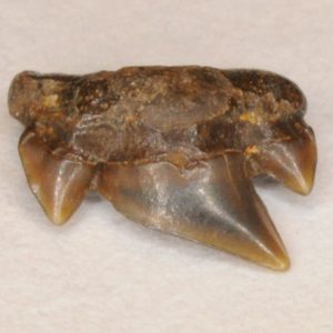
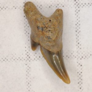
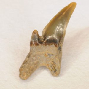
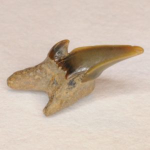
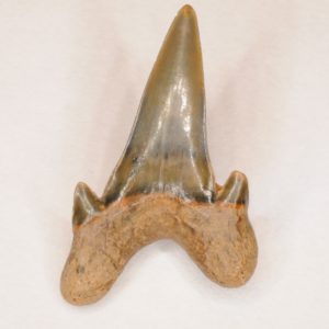
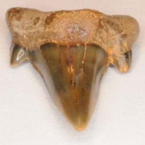
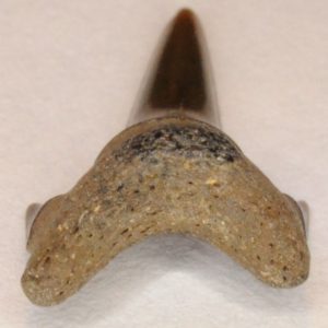
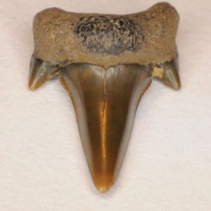
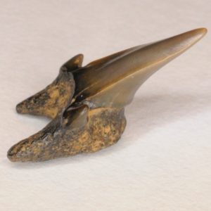
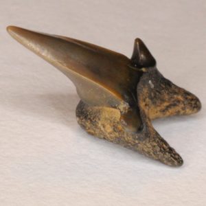
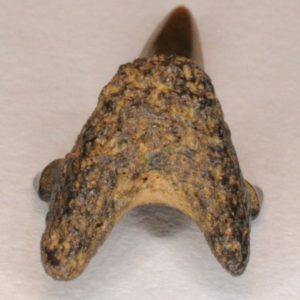
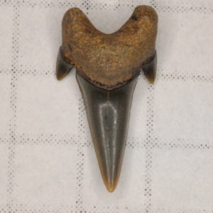
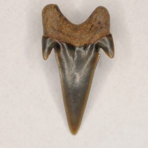
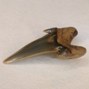
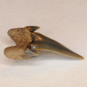
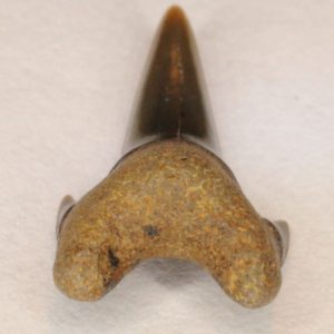
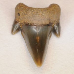
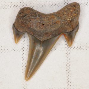
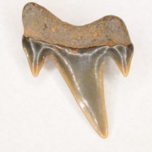
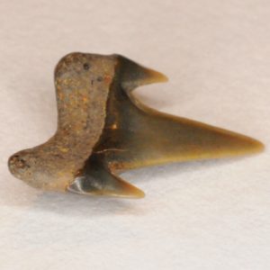
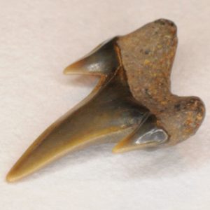
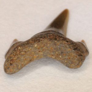
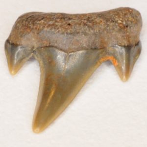
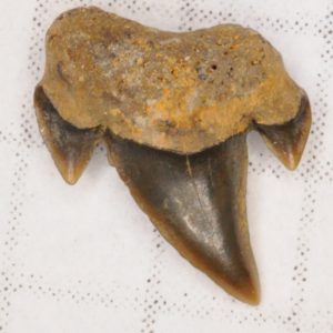
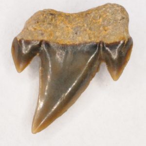
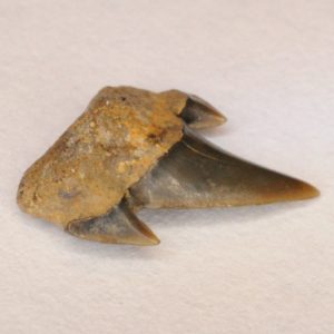
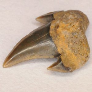
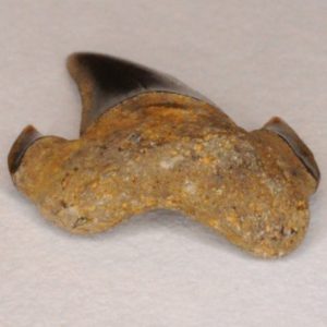
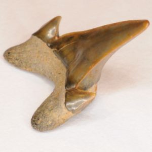
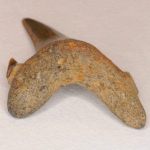
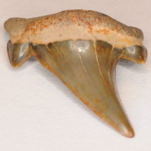
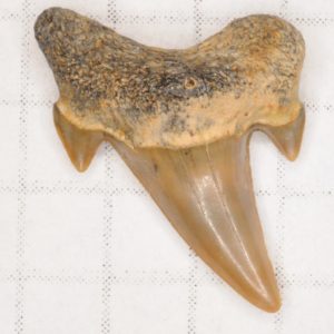
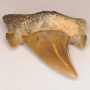
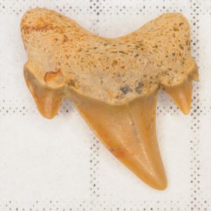
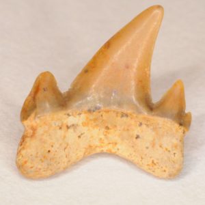
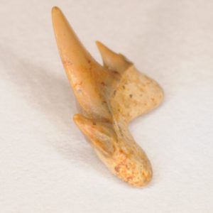
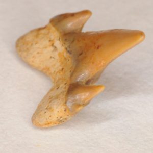
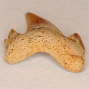
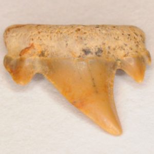
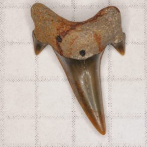
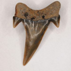
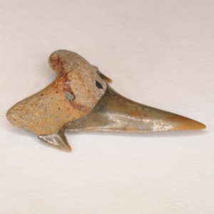
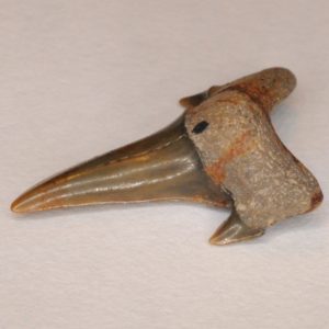
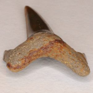
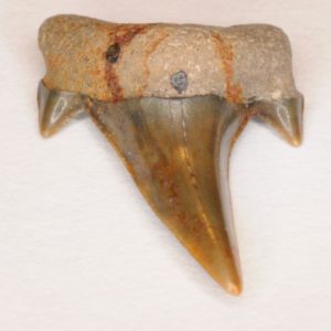
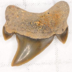
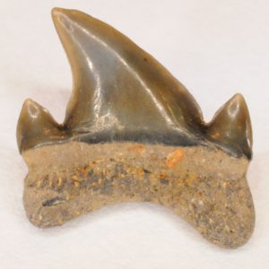
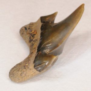
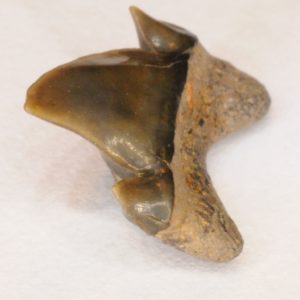
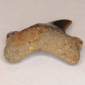
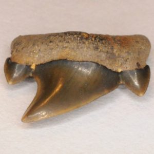
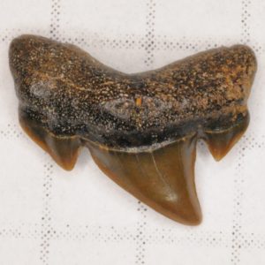
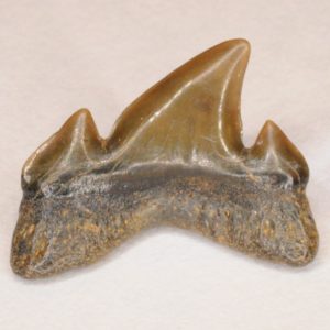
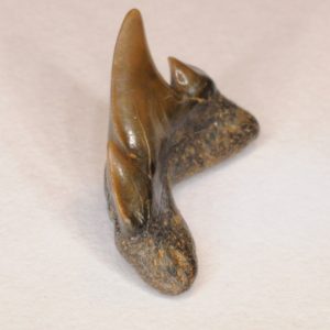
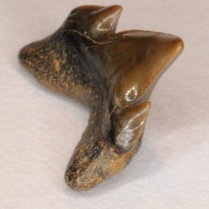
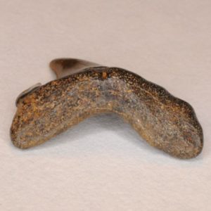
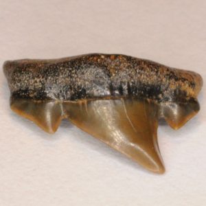
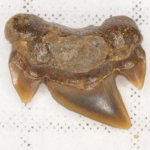
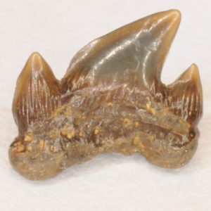
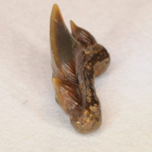
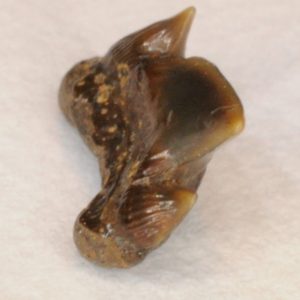
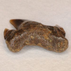
These teeth have a distinct look even when they are worn. They have a relatively robust triangular crown. They normally have a robust triangular cusplet on each side of the crown. The cusplets are rather massive for the size of the teeth and are thick at the base. They can sometimes have little secondary cusplets. The roots are bilobate. The lingual protuberance on these teeth juts out well on anterior teeth and diminishes a little in laterals and posteriors. The nutrient groove is missing on these teeth; instead, these teeth possess a nutrient pore. Some posterior teeth have ridges on the bottom of the labial face (these may not be present in all teeth). Laterals and posteriors are more curved distally than anteriors. This shark likely possessed two upper and one lower symphyseal/parasymphyseal teeth. It also had small intermediate teeth. The anteriors and laterals are enlarged in comparison to teeth of other positions. This shark had a crushing type of dentition.
Symphyseal / Parasymphyseal Teeth
Lingual View
Labial View
Profile View of Mesial Side
Profile View of Distal Side
Basal View
Occlusal View
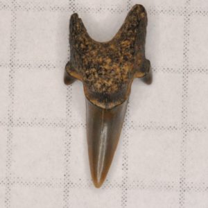
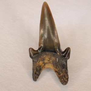
Symphyseal A. kopingensis teeth are harder to find because of their size. These teeth have a unique slender shape, almost resembling a sand tiger.
Anterior Teeth
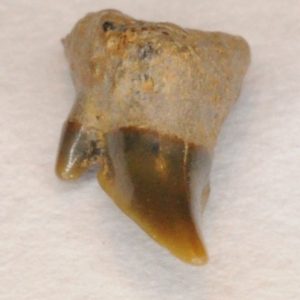
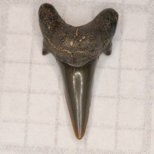
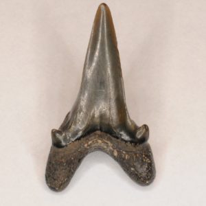
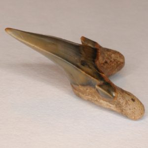
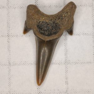
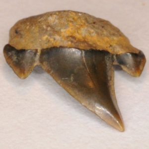
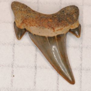
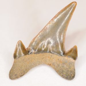
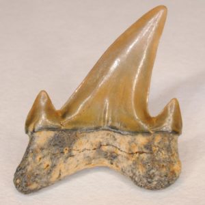
Lingual View
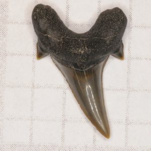
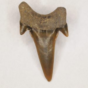
Labial View
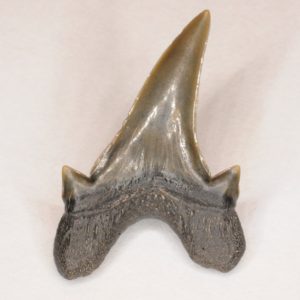
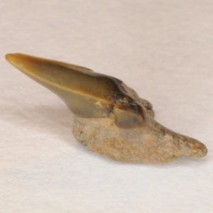
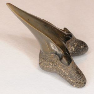
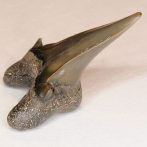
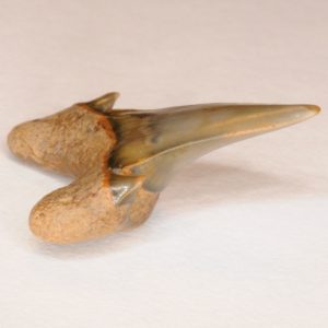
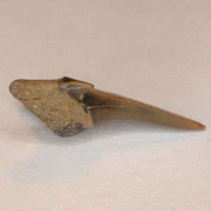
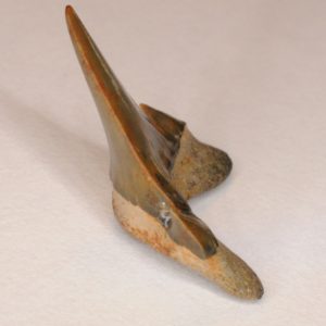
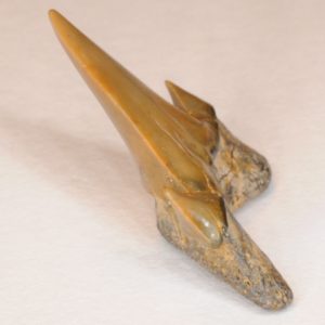
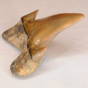
Profile View of Mesial Side
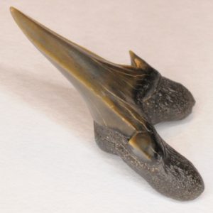
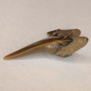
Profile View of Distal Side

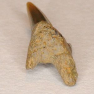
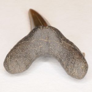
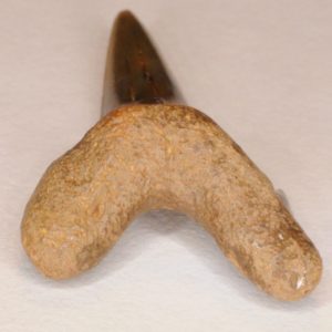
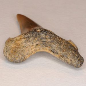
Basal View
Occlusal View
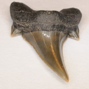
Lingual View
Labial View
Profile View of Mesial Side
Profile View of Distal Side
Basal View
Occlusal View
Lingual View
Labial View
Profile View of Mesial Side
Profile View of Distal Side
Basal View
Occlusal View
Lingual View
Labial View
Profile View of Mesial Side
Profile View of Distal Side
Basal View
Occlusal View
Lingual View
Labial View
Profile View of Mesial Side
Profile View of Distal Side
Basal View
Occlusal View

Lingual View
Labial View
Profile View of Mesial Side
Profile View of Distal Side
Basal View
Occlusal View
Intermediate Teeth
Lingual View
Labial View
Profile View of Mesial Side
Profile View of Distal Side
Basal View
Occlusal View
Lingual View
Labial View
Profile View of Mesial Side
Profile View of Distal Side
Basal View
Occlusal View
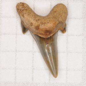
Lateral Teeth
Lingual View
Labial View
Profile View of Mesial Side
Profile View of Distal Side
Basal View
Occlusal View
Lingual View
Labial View
Profile View of Mesial Side
Profile View of Distal Side
Basal View
Occlusal View
Lingual View
Labial View
Profile View of Mesial Side
Profile View of Distal Side
Basal View
Occlusal View
Lingual View
Labial View
Profile View of Mesial Side
Profile View of Distal Side
Basal View
Occlusal View
Lower lateral teeth are not as curved and are less robust than the upper laterals. Lower lateral teeth are similar to lower anteriors, but are less robust and normally have a less prominent lingual protuberance. They also have a wider and “smoother” angle between the root lobes; lower anteriors have a narrow and “sharper” angle between the root lobes.
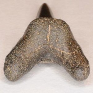
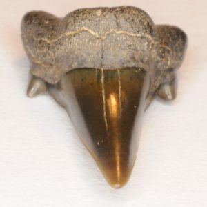
Posterior Teeth
Lingual View
Labial View
Profile View of Mesial Side
Profile View of Distal Side
Basal View
Occlusal View
Lingual View
Labial View
Profile View of Mesial Side
Profile View of Distal Side
Basal View
Occlusal View
Lingual View
Labial View
Profile View of Mesial Side
Profile View of Distal Side
Basal View
Occlusal View